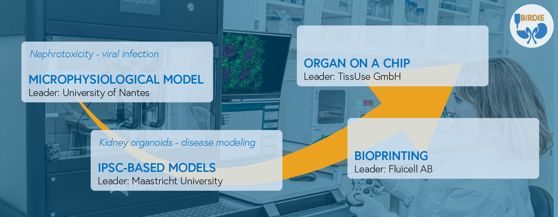
Microphysiological Model
Building up a biologically-relevant microphysiological model of the human renal tubulointerstitium is a complex task that requires to start with models of lower complexity. During BIRDIE project we are looking to set up long-term microphysiological models of human renal proximal cells cultured under physiological and disease states. Starting with a basic microphysiological model of human renal proximal cell monolayer using off-the-shelf materials from TissUse GmbH, this flat model will serve to define steady-state culture conditions under fluid shear stress, i.e., cell types, ECM component coating, culture media, etc. The optimized model will be challenged with both BK virus infection1,2,3 and nephrotoxic drugs, and transcriptomics will be used as a guidance system to constantly assess the proximity of our models compared to healthy or diseased native renal sorted cells and tissues.
iPSC-based models
Induced pluripotent stem cells (iPSCs) allow the generation of renal progenitors and organoids relevant for kidney in vitro models.6,7 Several models have been developed based on iPSCs-derived kidney organoids showing a certain degree of function of early rudiments. These self-assembled rudiments of the organ counterpart lack essential structures such as vasculature, which impairs the maturation and functions to the levels of the mature organ. IPSCs-derived organoids will be generated and combined with bioprinting and microfluidics for nephrotoxicity and viral infection screenings.
Bioprinting
Bioprinting technologies have gradually allowed to achieve an enormous progress on the manufacturing of tissue and organ-like 3D models, however these still display limited functionality.4 The combination of multiple bioprinting techniques is of paramount importance to achieve a broader range of complexity and mimicry in kidney models. Within BIRDIE we will use multiple bioprinting techniques to produce macro-size features and Fluicell’s unique single cell bioprinting5 to fine-tune cellular compositions within the kidney tubulointerstitium space.
Organ on a chip
Organ-on-a-chip systems enable a co-culture of physiologically relevant tissue models in a closed microfluidic circuit emulating the blood perfusion. Various multi-organ co-cultures have been performed previously on-chip and include liver8, 9, skin8, intestinal8, 9, neuronal9, kidney9, bone marrow10, vasculature11, pancreatic islet12, testis13 and lung14 models. The ability of the chips to host three-dimensional organ models in a controlled microenvironment under constant media perfusion enables them to create and maintain homeostasis. During the BIRDIE project a novel chip enabling a dual perfusion of a kidney model by overlapping a blood and a urinary microfluidic circuit will be developed by TissUse GmbH. This chip will harbor the 3D bioprinted models developed within the other work packages.
Scientific publications
- Maaike F.J. Fransen, Gabriele Addario, Carlijn V.C. Bouten, Franck Halary, Lorenzo Moroni, Carlos Mota. Bioprinting of kidney in vitro models: cells, biomaterials, and manufacturing techniques. Essays Biochem 2021; 65 (3): 587–602. doi: https://doi.org/10.1042/EBC20200158
- Francesca Perin, Eugenia Spessot, Anna Famà, Alessio Bucciarelli, Emanuela Callone, Carlos Mota, Antonella Motta, and Devid Maniglio. Modeling a Dynamic Printability Window on Polysaccharide Blend Inks for Extrusion Bioprinting. ACS Biomater. Sci. Eng. 2023, 9, 3, 1320–1331. https://doi.org/10.1021/acsbiomaterials.2c01143
- Michelle Lechtenberg, Coraline Chéneau, Kevin Riquin, Leopold Koenig, Carlos Mota, Franck Halary, Eva-Maria Dehne. A perfused iPSC-derived proximal tubule model for predicting drug-induced kidney injury. Toxicology in Vitro. 2025, 105. https://doi.org/10.1016/j.tiv.2025.106038
- Francesca Perin et al. Bioprinting of Alginate-Norbornene bioinks to create a versatile platform for kidney in vitro modeling. Bioactive Materials. 2025, 49. https://doi.org/10.1016/j.bioactmat.2025.03.010
Inventions recognized by the European Commission Innovation Radar
- New strategy to create thin channels with a microfluidic chip.
- Novel on-chip platform for 3D bioprinted renal model
- Integration of holographic phase microscopy (label-free imaging) as a quality control (QC) tool for bioprinting and tissue development
- BK virus infection diagnostics platform on new biomarkers
- Biopixlar Switching Head
White papers and application notes
- BIRDIE project and stakeholder report 2022 (Download PDF)
- BIRDIE project and stakeholder report 2023 (Download PDF)
References
- Sikorski M, et al. Non-permissive human conventional CD1c+ dendritic cells enable trans-infection of human primary renal tubular epithelial cells and protect BK polyomavirus from neutralization. PLoS Pathog. 2021; 17(2):e1009042. doi: 10.1371/journal.ppat.1009042.
- Masset C, et al. Resurgence of BK virus following Covid-19 in kidney transplant recipients. Transpl Infect Dis. 2021; 23(1):e13465. doi: 10.1111/tid.13465.
- McIlroy D, et al. Persistent BK Polyomavirus Viruria is Associated with Accumulation of VP1 Mutations and Neutralization Escape. Viruses. 2020; 12(8):824. doi: 10.3390/v12080824.
- Mota C, et al. Bioprinting: From Tissue and Organ Development to in Vitro Models. Chem Rev. 2020; 120(19):10547-10607. doi: 10.1021/acs.chemrev.9b00789.
- Jeffries GDM, et al. 3D micro-organisation printing of mammalian cells to generate biological tissues. Sci Rep. 2020; 10(1):19529. doi: 10.1038/s41598-020-74191-w.
- Wang S, Gao D, Chen Y. The potential of organoids in urological cancer research. Nat Rev Urol. 2017; 14(7):401-414. doi: 10.1038/nrurol.2017.65.
- Little MH and Combes AN. Kidney organoids: accurate models or fortunate accidents. Genes Dev. 2019; 33(19-20):1319-1345. doi: 10.1101/gad.329573.119.
- Maschmeyer I, et al. Chip-based human liver-intestine and liver-skin co-cultures–A first step toward systemic repeated dose substance testing in vitro. Eur J Pharm Biopharm. 2015; 95(Pt A):77-87. doi: 10.1016/j.ejpb.2015.03.002.
- Ramme AP, et al. Autologous induced pluripotent stem cell-derived four-organ-chip. Future Sci OA. 2019; 5(8):FSO413. doi: 10.2144/fsoa-2019-0065.
- Sieber S, et al. Bone marrow-on-a-chip: Long-term culture of human haematopoietic stem cells in a three-dimensional microfluidic environment. J Tissue Eng Regen Med. 2018; 12(2):479-489. doi: 10.1002/term.2507.
- Hasenberg T, et al. Emulating human microcapillaries in a multi-organ-chip platform. J Biotechnol. 2015; 216:1-10. doi: 10.1016/j.jbiotec.2015.09.038.
- Bauer S, et al. Functional coupling of human pancreatic islets and liver spheroids on-a-chip: Towards a novel human ex vivo type 2 diabetes model. Sci Rep. 2017; 7(1):14620. doi: 10.1038/s41598-017-14815-w.
- Baert Y, et al. A multi-organ-chip co-culture of liver and testis equivalents: a first step toward a systemic male reprotoxicity model. Hum Reprod. 2020; 35(5):1029-1044. doi: 10.1093/humrep/deaa057.
- Schimek K, et al. Human multi-organ chip co-culture of bronchial lung culture and liver spheroids for substance exposure studies. Sci Rep. 2020; 10(1):7865. doi: 10.1038/s41598-020-64219-6.
